Visiting Professor Mitsuru Morimoto
To understand organ development and tissue regeneration, we investigate mouse embryos and adult lung stem cells. The respiratory system achieves efficient gas exchange through proper branching airways and cell distribution. Our research focuses on the molecular mechanisms underlying development and regeneration across the lifespan, from embryo to adulthood. Specifically, we study lung tissue maturation during late development and the neonatal period, as well as tissue regeneration in the adult body. By deciphering these fundamental processes, we aim to gain insights into organ development and regenerative potential for future advancements.
Research and Education
The lung has long been perceived as a non-regenerative organ. However, researchers have uncovered intricate repair and regeneration mechanisms orchestrated by tissue-resident stem cells. Notably, the molecular dynamics governing stem cells during regeneration exhibit parallels with developmental processes, instigating inquiries into the conserved mechanisms underpinning both phenomena and their implications for pathological conditions.
In our laboratory, we utilize mouse genetics and human pluripotent stem cells to investigate: 1) the principles underlying lung development during the fetal to neonatal stages, 2) the mechanisms of adult tissue regeneration mediated by stem cells, 3) the modeling of respiratory diseases, and 4) reconstructing 3D lung tissue from stem cells. To address these research areas, we employ techniques such as organoid culture, high-resolution live imaging, single-cell RNA-sequencing. Graduate students will decide their research themes based on their interests and the laboratory’s direction. Our lab activities not only the acquisition of experimental techniques but also a deeper understanding of developmental biology, tips for reading scientific papers, and enhancement of academic presentation skills.
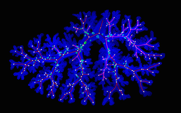 Figure 1. 3D-airway tissue structure of fetal mouse lung. The tissue is made transparent to visualize epithelial structure. Red lines indicate structures and white dots indicates branching points.
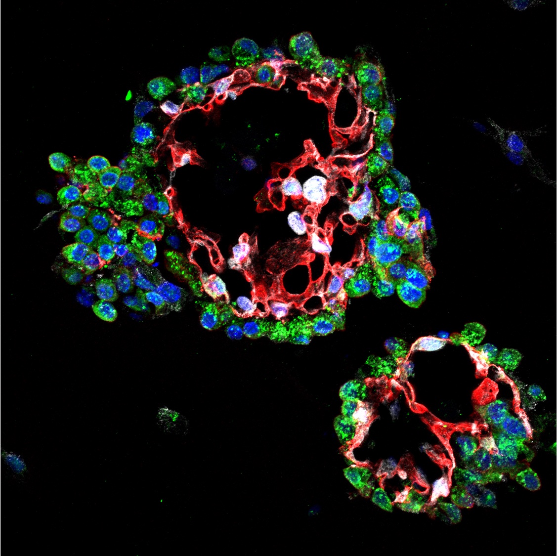 Figure 2. Sections of alveolar organoids. Alveolar organoids can be produced by 3D culture of tissue stem cells taken from lung tissue. Type I alveolar epithelial cells (red), type II alveolar epithelial cells (green).
Publications
- Hirofumi Kiyokawa, Akira Yamaoka, Chisa Matsuoka, Tomoko Tokuhara, Takaya Abe, Mitsuru Morimoto*. Airway basal stem cells reutilize the embryonic proliferation regulator, Tgfβ-Id2 axis, for tissue regeneration. Developmental Cell 56, 1917-1929 (2021).
- Kishimoto K, Furukawa KT, Luz-Madrigal A, Yamaoka A, Matsuoka C, Habu M, Alev C, Zorn AM, Morimoto M*. Bidirectional Wnt signaling between endoderm and mesoderm confers tracheal identity in mouse and human cells. Nature Communications vol. 11, Article number: 4159 (2020).
- Kishimoto K, Tamura M, Nishita M, Minami Y, Yamaoka A, Abe T, Shigeta M, Morimoto M*. Synchronized mesenchymal cell polarization and differentiation shape the formation of the murine trachea and esophagus. Nature Communications vol. 9, Article number: 2816 (2018).
- Tsao P*, Matsuoka C, Wei SC, Sato A, Sato S, Chena HK, Ling TY, Mori M, Cardoso WV, Morimoto M*. Epithelial Notch signaling regulates lung alveolar morphogenesis and airway epithelial integrity. Proc. Natl. Acad. Sci. USA 113, 8242-7 (2016)
- Noguchi M, Sumiyama K, Morimoto M*. Directed migration of pulmonary neuroendocrine cells toward airway branches organizes the stereotypic location of neuroepithelial bodies. Cell Reports 13, 2679–86 (2015)
Laboratory
[Visiting Professor] Mitsuru Morimoto
Yoshida-Konoe-cho, Sakyo-ku, Kyoto 606-8501, JAPAN
TEL: 075-753-4332
e-mail: mitsuru.morimoto@riken.jp