Ph.D. Associate Professor Tomohiro Ueno
In living organisms, biological activities are maintained based on various networks of biological organization. In our group, various imaging modalities such as MR microscopy are utilized to visualize individual living organisms in vivo with a high spatial resolution and ultimately to clarify those biological activities and disease progression. Especially, small animal models such as medaka are employed to obtain longitudinal changes in the same individual by in vivo time-series imaging. To accomplish these goals, our group is working on development of an imaging system, image analyses and animal disease models.
Research and Education
To visualize interactions in biological networks of an animal disease model, our group is working on development of an in vivo imaging system with high spatial resolution. As the imaging system, MR microscopy has been developed to be utilized in the same way as an optical microscope by increasing a spatial resolution of a noninvasive MRI. As the animal disease model, the medaka fish is employed. To achieve in vivo time-series imaging, its handling process such as an anesthesia procedure and an individual identification method has been also developed. As a result, fatty liver and cancer models of medaka have been visualized in vivo in a quantitative time-series way by using a hypothermic anesthesia method.
In our group, imaging principles and imaging hardware and software of the system that realize the principles are taught. In addition, group members are learning image analyses to extract characteristics of obtained images. Moreover, knowledge on mechanism of a disease and development of various animal disease models is obtained through research activities.
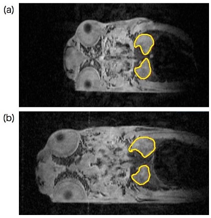 Time series coronal images of the same individual of a p53 knock-out medaka by the in vivo MR microscopy (a)10 weeks of age, (b) 14 weeks of age, outlined kidneys show asymmetrical changes
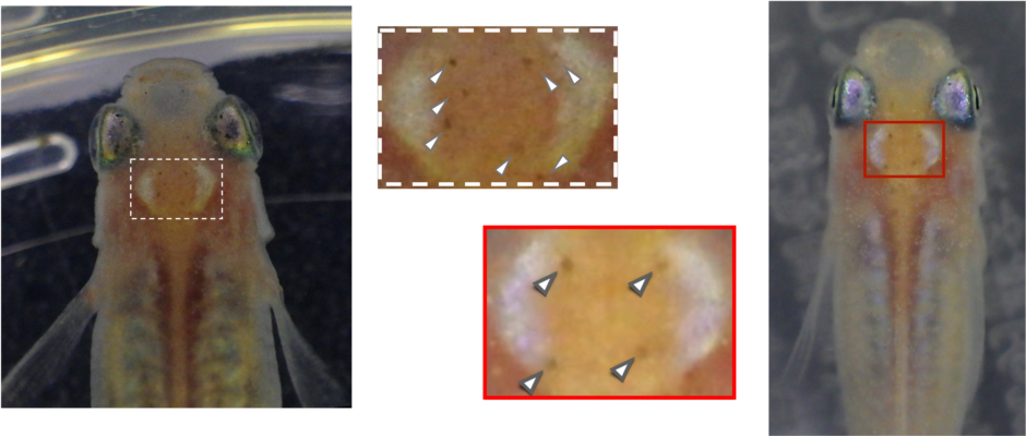 Individual identification of inbred medaka based on the melanophore spot patterns on the head(the different spot patterns appears in the red and white outlined regions)
Publication list
- Morizumi H, Sugimoto N, and Ueno T. “Individual identification of inbred medaka based on characteristic melanophore spot patterns on the head.” Sci. Rep. 13: 659 (2023).
- Watanabe S, Ueno T, Kimura Y, Mishina M, Sugimoto N. “Generative image transformer (GIT): unsupervised continuous image generative and transformable model for [123I]FP-CIT SPECT images” Ann. Nucl. Med. 35: 1203-1213 (2021).
- Suzuki T, Ueno T, Oishi N, Fukuyama H. “Intact in vivo visualization of telencephalic microvasculature in medaka using optical coherence tomography.” Sci. Rep. 10: 19831 (2020).
- Kishimoto K, Washio Y, Yoshiura Y, Toyoda A, Ueno T, Fukuyama H, Kato K, Kinoshita M. “Production of a breed of red sea bream Pagrus major with an increased skeletal muscle muss and reduced body length by genome editing with CRISPR/Cas9.” Aquaculture 495: 415-427 (2018).
- Ueno T, Suzuki H, Hiraishi M, Amano H, Fukuyama H, Sugimoto N. “In vivo Magnetic Resonance Microscopy and Hypothermic Anaesthesia of a Disease Model in Medaka.” Sci. Rep. 6: 27188; doi:10.1038/srep27188 (2016).
Laboratory
Tomohiro Ueno Associate Professor
Tel: +81-75-751-3907(office)
TEL: +81-75-751-3962(laboratory)
Email: ueno.tomohiro.2u@kyoto-u.ac.jp