Pathology is a school of medicine, which attempts to establish the basis of medical treatment by examining the difference between healthy and diseased tissues and thereby revealing the cause of diseases. Traditionally, we have been using macroscopic and microscopic methods with the diseased organs and tissues. However, we now adds not only new methodologies of molecular biology and genetics, but also state-of-art fluorescent imaging techniques, computer simulation, and mathematical modeling to challenge the questions of cancer and many other diseases.
Research and Education
The principal concept of our laboratory is “Cancer research based on microscopic imaging”. In 2001 we for the first time developed a probe for the activity of the Ras oncogene product, and thereafter we have developed nearly fifty probes for many signaling molecules. With several sophisticated microscopes such as multi-photon excitation microscopes, we are attempting to understand the disease by live-imaging of the very moment when diseases occur. In parallel, based on the multi-dimensional imaging data that contain the spatio-temporal information of molecular activities, we are building mathematical models of oncogene networks. Thus, we are moving forward from the classical pathology based on the observation of diseased tissues to pathology that live-images the disease in situ and that foresees the disease by simulation. Students in our laboratory come from schools of medicine, biology, engineering, physics etc., and study together on molecular biology, pathology, imaging technologies, systems biology etc. We speak English in the laboratory meetings to foster students who seek their future in the world.
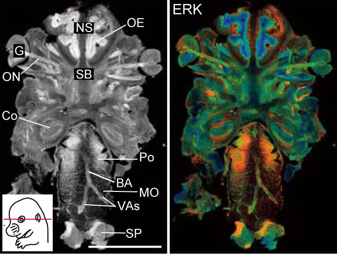 ERK activity map in a mouse
 Laboratory members
Recent Publications
- Ichise, H., Tsukamoto, S., Hirashima, T., Konishi, Y., Oki, C., Tsukiji, S., Iwano, S., Miyawaki, A., Sumiyama, K., Terai, K., and Matsda, M. Functional visualization of NK Cell-mediated killing of metastatic single tumor cells. eLife 11, e76269. (2022)
- Imanishi, A., Ichise, H., Fan, C., Nakagawa, Y., Kuwahara, K., Sumiyama, K., Matsuda, M., and Terai, K. Visualization of spatially controlled vasospasm by sympathetic nerve-mediated ROCK activation. Am J Pathol 161, 194-203. (2021)
- Konishi, Y., Ichise, H., Watabe, T., Oki, C., Tsukiji, S., Hamazaki, Y., Murakawa, Y., Takaori-Kondo, A., Terai, K., and Matsuda, M. Intravital imaging identifies the VEGF-TXA2 axis as a critical promoter of PGE2 secretion from tumor cells and immune evasion. Cancer Res 81, 4124-4132. (2021)
- Sato, T. Yamashita, M. Matsuda, Rhodopsin-mediated light-off-induced protein kinase A activation in mouse rod photoreceptor cells. Proc. Natl. Acad. Sci. U. S. A. 10.1073/pnas.2009164117 (2020).
- Kinjo, T., Terai, K., Horita, S., Nomura, N., Sumiyama, K., Togashi, K., Iwata, S., and Matsuda, M. FRET-assisted photoactivation of flavoproteins for in vivo two-photon optogenetics. Nat Methods. 16:1029-1036. (2019)