Ph.D.(in Eng.) Professor Naozo Sugimoto
Among biomedical imaging technologies, image information processing applicable to various clinical applications and ultra high resolution MRI are focused in this group. Medical image data obtained from such as CT and MRI becomes larger so as to provide medical staff with higher quality images. One of our objectives is to develop image information processing algorithm and systems to assist doctors in extracting necessary information from the huge data by more precise and more effective way. In order to obtain molecular/cell level information, we are also developing the MR microscopy system.
Research and Education
Two of ongoing research themes are introduced here.
We develop imaging and image processing techniques for quantitative analysis of multi dimensional medical images including multi modality images or sequential images. We also develop medical applications such as image guided radiotherapy, image guided intervention, and CAD(computer aided diagnosis) systems. Examples of the work are shown in the figures(Imaging and image processing for 4-dimensional cardiac MR tagged images, and MR DWI thermometry).
We have been also developing an ultra high resolution Magnetic Resonance Imaging(MRI), “MR microscopy”. With the MR microscope we have achieved a 20 um spatial resolution in small fresh-water fish, Medaka. Medaka have attracted many attentions as a cost-effective disease model. Since various physical properties can be used as contrast mechanism in MRI, we are trying to specify tissue/cell characteristics of medaka disease models in vivo not only from anatomical information but also from the physical properties. We have started applying quantitative susceptibility mapping to the MR microscope in order to quantify tissue iron contents.
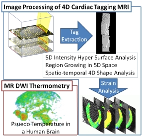 Multi dimensional image processing
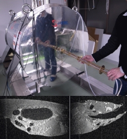 MR microscopy and MR microscopy images of medaka fish
Publications
- H. Morizumi, N. Sugimoto, and T. Ueno. Individual identification of inbred medaka based on characteristic melanophore spot patterns on the head, Sci. Rep. 13: 659 (2023), doi: 10.1038/s41598-023-27386-w
- S. Watanabe, T. Ueno., Y. Kimura, M. Mishina, N. Sugimoto. Generative image transformer (GIT): unsupervised continuous image generative and transformable model for [123I]FP-CIT SPECT images. Ann Nucl Med Vol.35, pp.1203-1213(2021). doi: 10.1007/s12149-021-01661-0
- I. Terada, Y. Togoe, T. Teratoko, T. Ueno, K. Ishizu, Y. Fujii, T. Shiina, N. Sugimoto, Monitoring of Portal Vein by Three-dimensional Ultrasound Image Tracking and Registration: Toward Hands-free Monitoring of Internal Organs , Advanced Biomedical Engineering , Vol.9, pp.1-9(2020), doi: 10.14326/abe.9.1
- M. Sasaki, M. Nakamura, T. Ono, R. Ashida, M. Yoshimura, M. Nakata, T. Mizowaki, N. Sugimoto, Positional repeatability and variation in internal and external markers during volumetric-modulated arc therapy under end-exhalation breath-hold conditions for pancreatic cancer patients, J. Radiat. Res., Vol.61, pp.755-765(2020), doi: 10.1093/jrr/rraa054
- H. Morizumi, T. Nishigaki, N. Sugimoto, T. Ueno, Individual Time Series Analysis of p53 Knockout Medaka by in vivo Magnetic Resonance Microscopy, JOINT ANNUAL MEETING ISMRM-ESMRMB(2018.June, Paris, France)
Laboratory
Professor: Naozo Sugimoto
TEL 075-751-4993
FAX 075-751-3909
e-mail sugimoto.naozo.8x@kyoto-u.ac.jp