M.D., Ph.D. Professor Tetsuya Takakuwa
Many people – some scientists, others not – are interested in early human development because it is closely related to the basic and important question: What are we and where have we come from? In our laboratory, morphogenesis during human embryonic and early-fetal period has been analysed using digitized datasets include high-resolution MRI, phase-contrast X-ray CT, and digitized histological serial sections obtained from the Kyoto Collection to reveal normal development as well as to elucidate the causes and pathogenesis of birth defects and to attempt to identify some preventive measures.
Research and Education
Human embryonic and early-fetal period has been analyzed three-dimensionally using digitized datasets. The digital data have gross merits that enabled us to develop various analyses, which accelerate the speed of morphological observations using precise and improve methods by providing a suitable plane for a morphometric analysis from many specimens. Morphometric data are useful for quantitatively evaluating and demonstrating the features of development and for screening abnormal samples, which may be suggestive in the pathogenesis of congenital malformations. The 3D coordinates of anatomical landmarks is provided for analyzing the positional change of interested landmarks and their relationships, and for mathematical analysis as well as statistical analysis such as Procrustes analysis and principle component analysis. Our 3D analysis may serve to provide accurate morphologic data, including the dynamics of embryonic structures related to developmental stages, which is required for insights into the dynamic and complex processes occurring during organogenesis. Our study could aid in differentiating normal and abnormal development, which may lead to prenatal diagnosis in the embryonic and early fetal period.
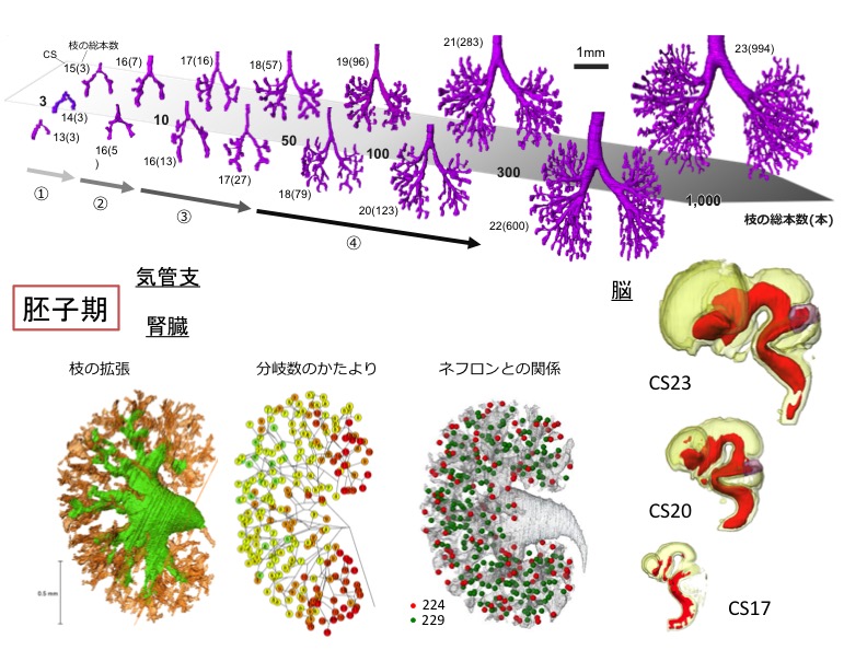 胚子期の器官形成(気管支、脳、腎臓)
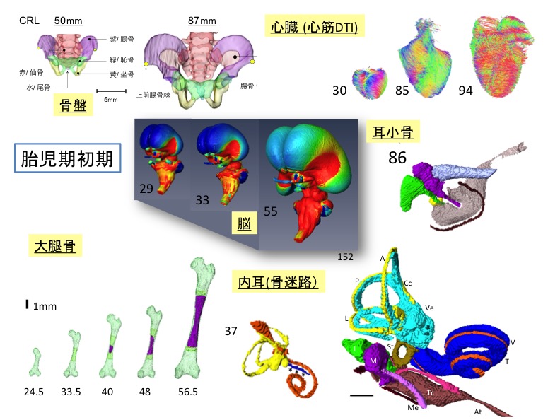 胎児期の器官形成(脳、骨盤、心臓、耳小骨、骨迷路、大腿骨)
Publications
- Iwasa Y, Kanahashi T, Imai H, Otani H, Yamada S, Takakuwa T. Human trapezius muscle development during early fetal period. J Anatomy 2024, in press, doi: 10.1111/joa.14116
- Kanahashi T, Matsubayashi J, Imai H, Yamada S, Otani H, Takakuwa T. Sexual dimorphism of the human fetal pelvis exists at the onset of primary ossification, Communications Biology, 2024, 7:538,https://doi.org/10.1038/s42003-024-06156-y
- Saizonou MA, Kitazawa H, Kanahashi T, Yamada S, Takakuwa T, Epithelial development of the urinary collecting system in the human embryo, PLOS ONE 19(4): e0301778. https://doi.org/10.1371/journal.pone.0301778
- Fujii S, Muranaka T, Matsubayashi J, Yamada S, Yoneyama A, Takakuwa T. Bronchial tree of the human embryo: Examination based on a mammalian model. J Anatomy 2024, 244(1), 159-169. http://doi.org/10.1111/joa.13946
- Fukui N, Kanahashi T, Matsubayashi J, Imai H, Yoneyama A, Otani H, Yamada S, Takakuwa T. Morphogenesis of the pulmonary vein and left atrial appendage in human embryos and early fetuses. J Anatomy 2024(1), 142-155, https://doi.org/10.1111/joa.13941
Laboratory
Professor: Tetsuya Takakuwa MD, PhD
Assistant Professor: Toru Kanahashi PhD
Human Health Science, Graduate School of Medicine, Kyoto University
606-8507 Sakyo-ku Shogoin Kawahara-cyo 53, Kyoto, Japan
TEL 075-751-3931/3956
e-mail; takakuwa.tetsuya.8x@kyoto-u.ac.jp
URL:http://pathology.hs.med.kyoto-u.ac.jp/